FREE! Click here to Join FunTrivia. Thousands of games, quizzes, and lots more!
Each Breath We take Trivia Quiz
Lung Volumes in Health and Disease
The amount of air we breathe with each breath is a small percentage of the air in our lungs. There are various parameters with the volume of air in the lungs that changes in both health and disease. Let's explore these different respiratory parameters.
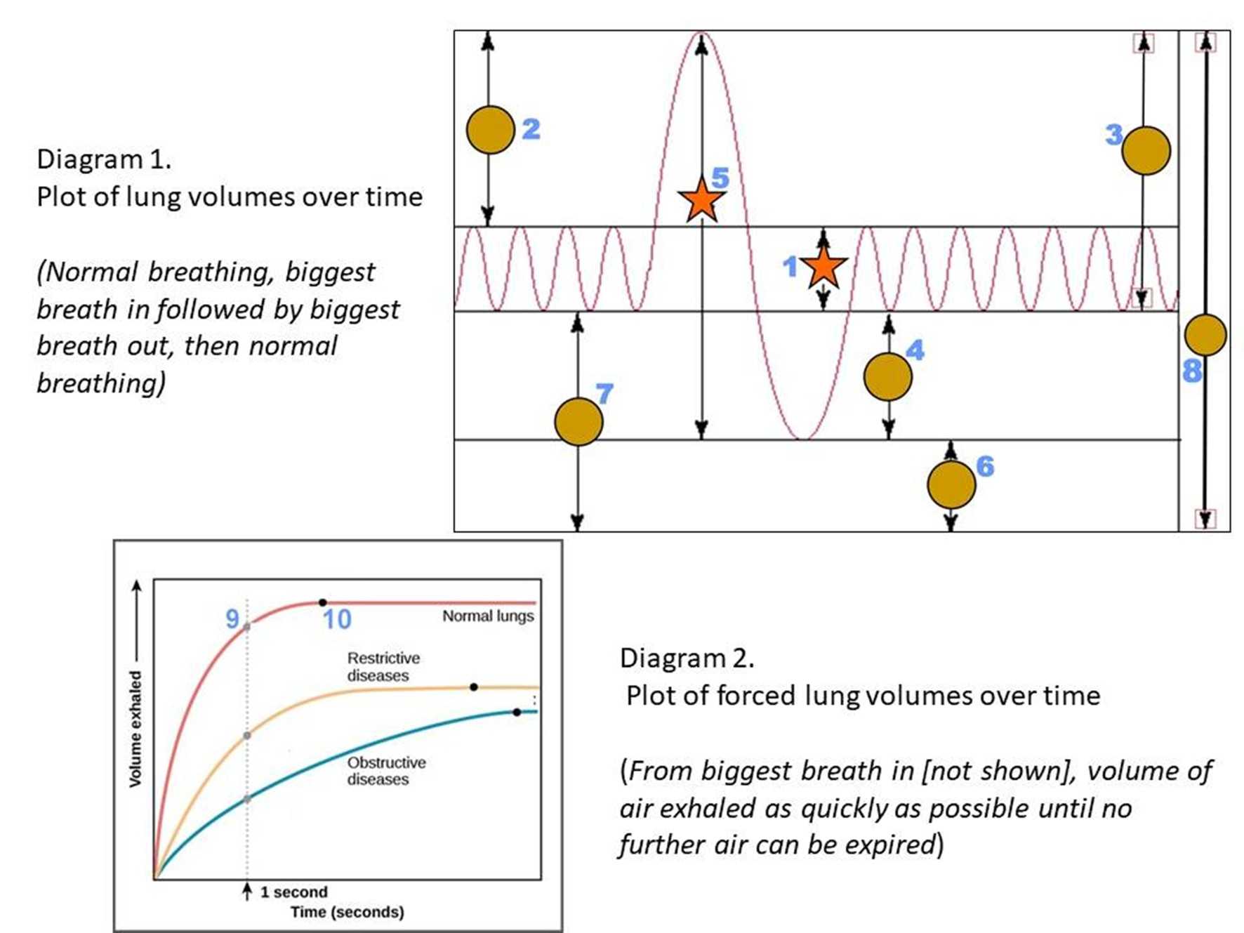
Diagrams 1 and 2 show plots of volumes of air over time. Diagram 1 shows quiet breathing followed by a deep breath blown all the way out. Diagram 2 shows, after a deep breath, the volume of air forced out of the lungs over time. Please match the lung volume parameter to each number.
Forced Expiratory Volume 1Tidal volumeVital capacityTotal Lung CapacityExpiratory Reserve VolumeFunctional Residual CapacityForced Vital CapacityInspiratory Reserve VolumeInspiratory CapacityResidual Volume* Drag / drop or click on the choices above to move them to the answer list.
1. Normal Breathing
2. Biggest breath in
3. Breath out, now *biggest breath in*
4. Breathe out as much as you can
5. Usable lung volume
6. Inaccessible air
7. Reserves of air
8.
9. Expiration as fast as possible
10. Very similar to another parameter
Most Recent Scores
Nov 12 2024
:
Guest 82: 2/10
Oct 30 2024
:
twlmy: 10/10
Oct 18 2024
:
Guest 207: 7/10
Oct 07 2024
:
gracie3: 2/10
Oct 05 2024
:
klotzplate: 10/10
Quiz Answer Key and Fun Facts
1. Tidal volume
Tidal Volume (TV) is the amount of air that can be inhaled or exhaled during one respiratory cycle. In normal adults, this is about 500mL which is a bit less than 10% of the total air in our lungs. Measurement of TV demonstrates the functions of the respiratory centre (in the brain), respiratory muscles and the mechanics of the lung and chest wall. TV is around 10% of the total air in the lungs showing we have plenty of reserves. A baby may breathe up to 30 times a minute at rest but an adult will only breathe 12-15 times a minute at rest.
Most (but not all lung volume measurements) can be made by connecting a patient to a spirometer which measures the amount of air displaced over time (Diagrams 1 and 2).
2. Inspiratory Reserve Volume
Inspiratory Reserve Volume (IRV) is the amount of air that can be forcibly inhaled AFTER a normal tidal volume. IRV is usually kept as a reserve but is used up during deep breathing on exertion. The normal adult value is 1800-3200mL.
3. Inspiratory Capacity
Inspiratory capacity (IC) is the maximum volume of air that can be inhaled following a resting state. ("Breath out, now take the biggest breath in"). It is calculated from the sum of inspiratory reserve volume and tidal volume.
IC = IRV+TV
IRV changes with age, gender, weight, height and ethnicity, physical activity, and altitude. These factors must be taken into consideration when trying to differentiate physiological reasons from pathological reasons for IRV changes.
4. Expiratory Reserve Volume
Expiratory Reserve Volume (ERV) is the volume of air that can be exhaled by force after exhalation of normal tidal volume. The normal adult value is 700-1200ml. ERV is reduced with weight gain, diseases where fluid accumulates in the trunk (ascites) or after upper abdominal surgery.
5. Vital capacity
Vital Capacity (VC) is the total amount of air exhaled after maximal inhalation ("Biggest breath in and all the way out"). VC is about 4800mL in the average male. It varies according to age and body height/weight. It can be calculated by adding tidal volume, inspiratory reserve volume, and expiratory reserve volume.
VC = TV+IRV+ERV.
When a person is running at maximum capacity, for example, they are using close to their VC volume with every breath plus their frequency of breathing also increases.
VC demonstrates the ability to breathe deeply and cough, which, in turn, indicates inspiratory and expiratory muscle strength. VC should be 3 times greater than TV for an effective cough response. VC is always reduced in restrictive disorders and sometimes in obstructive diseases. (See Q10).
6. Residual Volume
Residual Volume (RV) is the volume of air remaining in the lungs after maximal exhalation. In a normal adult male this averages around 1200ml. It is indirectly measured by adding FRC (see Q7) and ERV, and requires more sophisticated measuring devices as it cannot be measured by spirometry.
Incomplete emptying of the lungs and air trapping is typical of obstructive lung diseases. RV, in these cases, will nearly always be high.
The RV can also be expressed as a percentage of total lung capacity and those values higher than 40% significantly increase the risks of pneumothorax (air in the chest cavity not lungs), and chest infection.
7. Functional Residual Capacity
Function Residual Capacity (FRC) is the amount of air remaining in the lungs at the end of a normal exhalation. It can be calculated by adding together residual and expiratory reserve volumes. The normal value in the adult male is about 1750 - 2250 mL or about one-third of total lung capacity. In effect, it is the amount of air reserves in the lungs
FRC = RV+ERV.
FRC unlike IC does not rely on effort and indicates the resting position of the lungs. FRC is decreased in restrictive disorders.
The ratio of FRC to TLC is a measure of hyperinflation (blockages in the air passages or by air sacs with less elasticity, which interferes with the exhalation of air from the lungs).
In Chronic Obstructive Pulmonary Disease, FRC is up to 80% of total lung capacity
8. Total Lung Capacity
Total Lung Capacity (TLC) is the maximum volume of air the lungs can hold. It is the sum total of all volume compartments (ie volume of air in lungs after a maximum inspiration). The normal value is about 6.0 L in adult males and 4.8 L in adult females. TLC is calculated by adding the four primary lung volumes.
TLC = TV + IRV + ERV + RV
TLC can be increased in patients with obstructive diseases such as emphysema. It can be decreased in patients with restrictive diseases such as chest wall abnormalities.
9. Forced Expiratory Volume 1
(Diagram 2) Forced expiratory volume in 1 second (FEV1) is the volume of air that can be blown out in the first second by FORCE, after one full inspiration. Average values for FEV1 in healthy people depend mainly on sex, race and age. There is a wide range of 'normal' values with values between 80% to 120% of the average value considered normal. Predicted normal values for FEV1 can be calculated and depend on age, sex, height, mass and ethnicity. Young adults can forcibly expire 80% of their Forced Vital Capacity in one second and this decreases normally with age to about 70%.
10. Forced Vital Capacity
To measure Forced Vital Capacity (FVC) by spirometer, the patient is directed to take the deepest breath possible and then exhale into the spirometer as fast as possible, for as long as possible, ideally for at least 6 seconds. (During the test, nose clips are used to prevent air from escaping through the nostrils). The accuracy of the test is highly dependent on patient cooperation and effort and is normally repeated to ensure reproducibility. FVC approximates VC but because of patient cooperation, FVC can only be underestimated and never overestimated. Nevertheless, in a compliant patient, the FVC is very close to the VC.
FEV1/FVC is the ratio of FEV1 to FVC. This should be approximately 70-80% (the parameter declines with age). Standardised tables are available to determine the correct value for any particular person based on height, race and gender.
In obstructive diseases (asthma, chronic bronchitis, and emphysema) FEV1 is reduced because of increased airway resistance to expired airflow. The FVC may be decreased as well, due to the premature closure of airways in expiration. However, will not be as marked as the reduction in FEV1 (for example, both FEV1 and FVC are decreased, but the FEV1 is more affected because of the increased airway resistance). This generates a reduced value which can be as low as 30% (Reference range 70-80%). To distinguish between asthma and chronic bronchitis which both present with similar symptoms, clinically, The FEV1/FVC test is performed and then a bronchodilator dose is given to the patient. After it has been allowed to 'work', the test is repeated. If the FEV1/FVC increases significantly, this is diagnostic of asthma. If there is no significant increase, then a diagnosis of chronic bronchitis is made.
In restrictive diseases (such as pulmonary fibrosis) the FEV1 and FVC are both reduced proportionally and the value of FEV1/FVC may be normal. However, the diagnostic parameter is the FVC which will always be much lower than the FVC of a healthy person.
This quiz was reviewed by FunTrivia editor WesleyCrusher before going online.
Any errors found in FunTrivia content are routinely corrected through our feedback system.