FREE! Click here to Join FunTrivia. Thousands of games, quizzes, and lots more!
Quiz about The Eyes Have It

10 Questions about The Eyes Have It | Sci / Tech


Can you match each of these parts of the human eye with its function? Let's see.

A matching quiz by looney_tunes. Estimated time: 4 mins.
  1. Home
  2. »
  3. Quizzes
  4. »
  5. Science Trivia
  6. »
  7. Nervous System and the Senses
  8. »
  9. Eyes and Vision

Author
looney_tunes
Time
4 mins
Type
Match Quiz
Quiz #
380,141
Updated
Dec 03 21
# Qns
10
Difficulty
Average
Avg Score
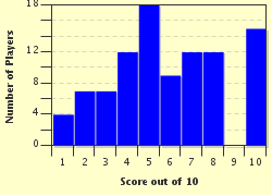
7 / 10
Plays
704
Awards
Top 20% Quiz
Last 3 plays: Guest 107 (10/10), Guest 47 (4/10), Guest 108 (10/10).
(a) Drag-and-drop from the right to the left, or (b) click on a right side answer box and then on a left side box to move it.
QuestionsChoices
1. White of the eye, protects and encloses the eye's contents.  
  vitreous humour
2. Jelly-like material forming most of the eye's volume.  
  sclera
3. Provides lubrication for the eye.  
  cornea
4. Contains blood vessels supplying the retina with oxygen and nutrients.  
  iris
5. Clear covering at front of the eye which allows light in.  
  conjunctiva
6. Ring of muscles that contract to control the amount of light entering the eye.  
  fovea
7. Adjusts shape to focus incoming light on the retina.  
  cone
8. Part of the retina that is most sensitive to light.  
  optic nerve
9. Type of retinal cell which is used in detecting colour.  
  choroidea
10. Transmits information from eye to brain.  
  lens


Select each answer

1. White of the eye, protects and encloses the eye's contents.
2. Jelly-like material forming most of the eye's volume.
3. Provides lubrication for the eye.
4. Contains blood vessels supplying the retina with oxygen and nutrients.
5. Clear covering at front of the eye which allows light in.
6. Ring of muscles that contract to control the amount of light entering the eye.
7. Adjusts shape to focus incoming light on the retina.
8. Part of the retina that is most sensitive to light.
9. Type of retinal cell which is used in detecting colour.
10. Transmits information from eye to brain.

Most Recent Scores
Nov 18 2024 : Guest 107: 10/10
Nov 17 2024 : Guest 47: 4/10
Nov 14 2024 : Guest 108: 10/10
Nov 07 2024 : twlmy: 10/10
Nov 02 2024 : bradez: 7/10
Oct 08 2024 : Guest 198: 0/10

Score Distribution

quiz
Quiz Answer Key and Fun Facts
1. White of the eye, protects and encloses the eye's contents.

Answer: sclera

The word sclera comes from a Greek word meaning hard - it is the tough outer layer of the back portion of the eyeball, made of collagen and elastic fibres so that it can offer protection and keep the globe of the eye in shape. It is also the part of the eye to which muscles attach, and has a number of tiny holes through which nerves and blood vessels pass.

The relatively small size of the iris makes the human 'white of the eye' a prominent feature. In many animals, it is not entirely white, but the visible portion is the same color as the iris. In horses, for example, you will only see the white of the eye if the animal is upset - it is standard advice to someone buying a first horse that they should avoid purchasing an animal who displays the whites of their eyes, as it is likely to be a bit difficult to handle for a novice owner/rider.
2. Jelly-like material forming most of the eye's volume.

Answer: vitreous humour

Also called the vitreous body, the vitreous humour is what makes the eye have a spherical shape. It is mostly water (98-99%), but the other cells and dissolved chemicals give it a viscosity ('thickness' or tendency to hold together rather than spreading out) that is similar to that of jelly. Vitreous humour does not replenish itself, so if any impurities enter it, they stay there unless surgically removed - if the particles are large enough to be seen and interfere with clarity of vision, they are called floaters.
3. Provides lubrication for the eye.

Answer: conjunctiva

The conjunctiva has three layers: the palpebral layers lines the eyelids, the bulbar layer covers the front part of the sclera, and the fornix layer joins the other two. The conjunctiva keeps the sclera moist through the production of mucus and tears (although it is the lacrimal gland that produces many more tears, as when your eyes are watering).

It also helps to keep microbes from entering the eye. It may get inflamed (a condition known as conjunctivitis, commonly called pinkeye), either because of infection or because of an autoimmune response.

The condition will often clear without requiring treatment, but antibiotics may be prescribed for a bacterial infection, or antihistamines for an allergic reaction.
4. Contains blood vessels supplying the retina with oxygen and nutrients.

Answer: choroidea

Also known as the choroid coat, or just the choroid, this is part of the uveal tract, the blood-carrying layer of the eye, between the sclera and the retina. (The iris and the ciliary body form the rest of the layer.) As well as providing nutrients, the choroid also helps the eye collect light efficiently - its black color reduces the amount of light that is reflected from its surface back into the interior of the eye.

It also absorbs light which comes through the sclera, thereby making the image formed on the retina clearer than it might otherwise be.
5. Clear covering at front of the eye which allows light in.

Answer: cornea

The cornea provides about two-thirds of the refraction that allows the eye to focus incoming light onto the retina. However, its refraction is fixed, so the actual fine-tuning is done by the lens. Because its task requires that it be transparent, the cornea does not contain any blood vessels, but gains essential nutrients primarily by absorbing them from tear fluid on its outer surface, and from the aqueous humor on its inner surface. Reshaping the cornea to reduce or eliminate the need for corrective devices (glasses or contact lenses) can be performed, most commonly using an exiplex laser. (I would rather wear glasses as I have for over fifty years than have someone fooling around with my eyes, but LASIK surgery is very popular.)
6. Ring of muscles that contract to control the amount of light entering the eye.

Answer: iris

The iris, which is seen as the coloured part of the eye, is annular in shape - the opening inside the circle of contractile fibres is called the pupil. There are two sets of muscles in the iris: the sphincter pupillae and the dilator pupillae. The former form a circular surround of the iris; their contraction makes the iris shrink inwards, and the pupil gets smaller in size. The latter are arranged in a radial formation; when they contract the pupil gets larger.

The colour of the iris depends on the relative amount of two types of melanin molecule: brown-eyed people have a higher ratio of eumalanin (brown or black) to pheomelanin (red) than is the case for those with blue or green eyes. Albinos have no melanin, and their eyes appear pink since there is no melanin color to cover the colour of the blood vessels in the eye.
7. Adjusts shape to focus incoming light on the retina.

Answer: lens

While most of the focusing of light is done by the cornea, and the aqueous and vitreous humours also contribute, it is the lens which can change shape to get a precise image for objects at varying distances. It is a biconvex lens (both sides bulge outwards when it is viewed from the side), and muscles attached to it can make it thinner (when they contract) to provide a clear image for distant objects, or thicker (when they relax) to provide a clear image for close objects.

The lens may become partially or completely opaque - a condition referred to as cataracts, which cause about half of the blindness in the world. They can be treated by replacing the lens with a synthetic replacement. They are most commonly associated with aging, as is presbyopia, a loss of the ability of the lens to change shape that leads to difficulty focusing on close objects.
8. Part of the retina that is most sensitive to light.

Answer: fovea

The retina is the layer of cells in the eye which contains light-sensitive cells to change incoming light signals to electrical impulses that can be sent to the brain and interpreted as images. Near the centre at the back of the eye is the macula lutea, an oval region which contains several regions of cells designed for light detection, including the fovea (or, to give it its full name, the fovea centralis).

It is a shallow indentation (hence its name, which means pit) in the macula, containing the highest concentration of cones of any region in the eye. About half of the nerve fibres in the optic nerve carry information from the fovea.
9. Type of retinal cell which is used in detecting colour.

Answer: cone

As well as providing fine detail for vision, the cones are the cells which respond to different wavelengths of light so as to allow us to see colours. The other type of light-detecting cells in the retina are called rods. Cones only operate effectively when there is ample illumination - in dim light, the rods still operate effectively, but the cones do not. This is why objects seem to lose colour when seen in dim light.

Because the fovea has only cones, you can improve your vision in dim light by looking out of the side of your eye, allowing the parts of the retina with rods to form the basis of your vision. This is a trick that is commonly used by optical astronomers, whose work is obviously done in dim light.
10. Transmits information from eye to brain.

Answer: optic nerve

The optic nerve is one of the twelve paired cranial nerves, nerves that come directly from the brain rather than the spinal cord, and appear in pairs, with one going to each side of the body. Unlike most of the cranial nerves, it is actually enclosed by the three meningeal layers that surround the brain, and is considered part of the central, rather than the peripheral, nervous system.

Because there are no light-detecting cells at the point where the optic nerve is attached to the retina, each eye has a blind spot - light falling there does not send an impulse to the brain. While both eyes are active, the other eye compensates for this small gap in your vision, but if you close one eye and move an object around while keeping your open eye focused on some distant point, you can see the moving object disappear as it passes through the blind spot.
Source: Author looney_tunes

This quiz was reviewed by FunTrivia editor rossian before going online.
Any errors found in FunTrivia content are routinely corrected through our feedback system.
Related Quizzes
This quiz is part of series The Joy of Science:

A collection of quizzes exploring some of the fascinating aspects of science, aimed at players with a high school science background. Or those who are just curious about their world.

  1. Heavy Metal Lover Easier
  2. The Physical Revue Featuring Tom Lehrer Average
  3. A Chemical Tourist Easier
  4. A Category of One's Own Average
  5. Nuclear Fission For Kids! Average
  6. Molecules of Life Average
  7. The Hertzsprung-Russell Diagram Average
  8. Signs for Designs Tough
  9. Billions and Billions of Stars Easier
  10. The Eyes Have It Average
  11. The Power is Yours Average
  12. Mohs Scale Easier

11/21/2024, Copyright 2024 FunTrivia, Inc. - Report an Error / Contact Us