FREE! Click here to Join FunTrivia. Thousands of games, quizzes, and lots more!
Quiz about Fifteen Minutes in the Cell

Fifteen Minutes in the Cell Trivia Quiz


You've read about cellular biology, but can you identify cells and structures from electron microscope images? In fifteen minutes, you'll know if you take this quiz. I'll give you some hints, too!

A photo quiz by WesleyCrusher. Estimated time: 6 mins.
  1. Home
  2. »
  3. Quizzes
  4. »
  5. Science Trivia
  6. »
  7. Biology
  8. »
  9. Cell and Molecular Biology

Time
6 mins
Type
Photo Quiz
Quiz #
335,907
Updated
Dec 03 21
# Qns
10
Difficulty
Tough
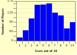
Avg Score
6 / 10
Plays
8386
Awards
Top 5% quiz!
Last 3 plays: Guest 82 (3/10), cluelesskat (5/10), Hando (4/10).
-
Question 1 of 10
1. Many cells are easily identified by their shapes. Here, we see some human cells. Even if you don't know what exactly they are, some thinking about the purpose of the cells should tell you these are which type? Hint


Question 2 of 10
2. Here, we see a point where three cells meet. An expert might recognize the species, but just from the look of the division between the cells, you can safely state that these are from which type of life? Hint


Question 3 of 10
3. The internal structures can also sometimes tell you a lot about a cell. In this case, one of these four structures is not something you would normally see in a cell at rest. Which one of them tells the cell sleuth something and what is the verdict? (Click on the image to zoom if you need a clearer picture) Hint


Question 4 of 10
4. Here we have a rather large cell organelle that clearly shows some subdivisions. It is also something a human cell usually has exactly one of. Which is it? Hint


Question 5 of 10
5. This organelle looks almost like a fingerprint but it is rather a network to which other, very small but important cell components attach while doing their function. Its name also suggests "little network" in Latin; which organelle is it? Hint


Question 6 of 10
6. This image belongs to the same cell as the one before, but here, there are black structures embedded. They are not attached to the network but just happen to be here because the network needs a lot of energy. What is the name of these organelles? Hint


Question 7 of 10
7. This is not a detached part of the cellular component seen in question 5, but a rather different organelle whose main function is to transport vesicles - capsules containing important chemicals - to their destination. What is its rather mechanical name? Hint


Question 8 of 10
8. Here we are looking at a rather rare perspective of one particular organelle that actually has the shape of a drinking straw. The photo shows one of its ends and its microstructure involving nine small bundles of what again looks like straws. The name of this organelle is misleading, considering it rather organizes the peripheral structure of the cell. What do you see here? Hint


Question 9 of 10
9. Uh-oh! Something is happening here! What is going on in this particular picture? The process is definitely more advantageous to the particles on the left than to the mass on the right. Hint


Question 10 of 10
10. Finally, let's take a look at some chromosomes. These three chromosome pairs have been taken from a human karyotype image. Which of the following statements about them is definitely WRONG, keeping in mind the convention for labeling chromosomes? Hint


(Optional) Create a Free FunTrivia ID to save the points you are about to earn:

arrow Select a User ID:
arrow Choose a Password:
arrow Your Email:


View Image Attributions for This Quiz

Most Recent Scores
Jan 25 2025 : Guest 82: 3/10
Jan 19 2025 : cluelesskat: 5/10
Jan 16 2025 : Hando: 4/10
Jan 15 2025 : Shiary: 4/10
Jan 14 2025 : violet71: 2/10
Jan 14 2025 : Guest 159: 3/10
Dec 28 2024 : mollybobbins: 6/10
Dec 19 2024 : Guest 67: 8/10
Dec 14 2024 : Guest 174: 8/10

Score Distribution

quiz
Quiz Answer Key and Fun Facts
1. Many cells are easily identified by their shapes. Here, we see some human cells. Even if you don't know what exactly they are, some thinking about the purpose of the cells should tell you these are which type?

Answer: Blood cells

Even at a glance, two features are quite apparent about these cells: They are not connected and they do not possess a means of locomotion. The first observation rules out muscle and nerve cells, both of which require a tight connection to do their job and the second rules out sperm which has a mobile tail (cilium) for movement. Thus, these are blood cells - the puffy balls are leukocytes (white blood cells) and the flat disks are erythrocytes (red blood cells).
2. Here, we see a point where three cells meet. An expert might recognize the species, but just from the look of the division between the cells, you can safely state that these are from which type of life?

Answer: A plant

These cells are divided from each other not only by a cell membrane, but also by a clearly visible, stiff, thick cell wall. Only plant cells possess this feature.
3. The internal structures can also sometimes tell you a lot about a cell. In this case, one of these four structures is not something you would normally see in a cell at rest. Which one of them tells the cell sleuth something and what is the verdict? (Click on the image to zoom if you need a clearer picture)

Answer: Structure 3 shows the cell is in the process of dividing.

The structure you see in number 3 is called the central plate, composed of the cell's already duplicated chromosomes lined up and ready for pair separation. All other components are normal organelles of a plant root cell, also already separating in preparation for a normal mitotic cell division.
4. Here we have a rather large cell organelle that clearly shows some subdivisions. It is also something a human cell usually has exactly one of. Which is it?

Answer: The nucleus

What we see here is the nucleus - the control center of the cell. Like all major organelles, it has a membrane of its own filled with plasma. In this plasma, the chromatin material (chromosomes in their unwound state) is floating. The dark spot in the middle of the nucleus is the nucleolus ("little core") where a particular type of RNA that controls cell activity is made. Cells usually only have one nucleus although there are some exceptions, such as in skeletal muscle cells.
5. This organelle looks almost like a fingerprint but it is rather a network to which other, very small but important cell components attach while doing their function. Its name also suggests "little network" in Latin; which organelle is it?

Answer: The endoplasmic reticulum

In most animal cells, the endoplasmic reticulum attaches directly to the nucleus. On its surface, there are many ribosomes attached whose job it is to synthesize proteins based on the blueprints in the messenger RNA sent from the nucleus. Once the proteins are completed, the endoplasmic reticulum assists in their correct folding and then they are transported to their final destination in the cell.
6. This image belongs to the same cell as the one before, but here, there are black structures embedded. They are not attached to the network but just happen to be here because the network needs a lot of energy. What is the name of these organelles?

Answer: Mitochondria

Mitochondria are the power plants of the cell. They control the most critical processes of cellular respiration, the process that breaks down glucose and oxygen into carbon dioxide and water. One molecule of glucose theoretically powers up 38 molecules of ATP, the universal intracellular energy carrier, but due to leaky membranes and the need for transporting reaction products, the net yield is about 28 to 30.

This process actually converts only one third of the original energy stored in the glucose into a usable form - not very efficient, but still 15 times more efficient than fermentation.
7. This is not a detached part of the cellular component seen in question 5, but a rather different organelle whose main function is to transport vesicles - capsules containing important chemicals - to their destination. What is its rather mechanical name?

Answer: Golgi apparatus

The Golgi apparatus is essentially a folded membrane on which the individual vesicles drift, powered towards their destination by chemical reactions along the surface. Many of the vesicles thus transported are actually small bubbles of material that detached itself from the endoplasmic reticulum or, more rarely, the Golgi apparatus itself.

They are then often reabsorbed at their destination.
8. Here we are looking at a rather rare perspective of one particular organelle that actually has the shape of a drinking straw. The photo shows one of its ends and its microstructure involving nine small bundles of what again looks like straws. The name of this organelle is misleading, considering it rather organizes the peripheral structure of the cell. What do you see here?

Answer: A centriole

Centrioles are the guides and architects of the microtubule structure that ultimately makes up the cytoskeleton of a cell. Besides giving the cell a more consistent shape, these structures are involved in cell division and transport. If a cell has a flagellum (a "tail" used for moving) or other means of locomotion, the centrioles and microtubuli control its action as well.
9. Uh-oh! Something is happening here! What is going on in this particular picture? The process is definitely more advantageous to the particles on the left than to the mass on the right.

Answer: HIV viruses are infecting a blood cell.

This is definitely something that you do not want to happen in your own body: What we see here are four HIV particles of which two have just started the infection process by docking to receptors on the cell surface. The cell will then actually engulf the virus and start dissolving it, releasing its genetic material to the cell plasma. From there, it can then undermine the cell's own molecular program and use the cell's resources to multiply.
10. Finally, let's take a look at some chromosomes. These three chromosome pairs have been taken from a human karyotype image. Which of the following statements about them is definitely WRONG, keeping in mind the convention for labeling chromosomes?

Answer: Pair C shows chromosome 22.

Pair B are unequal, marking them as an X and a Y chromosome, thus identifying a male. Pairs A and C are normal autosomes (standard paired chromosomes equal in both genders), but the numbering convention places the largest chromosomes first. Thus, C, clearly very large, cannot have a high number (in fact it is chromosome 2). Finally, we can neither prove nor disprove the genetic health of the individual.

There is nothing visibly wrong with these three pairs, but one of the other twenty could be defective or there could be a small, invisible but still fatal mutation present.
Source: Author WesleyCrusher

This quiz was reviewed by FunTrivia editor crisw before going online.
Any errors found in FunTrivia content are routinely corrected through our feedback system.
Related Quizzes
This quiz is part of series Commission #15:

You could very well work through these quizzes in a quarter of an hour if you really put your mind to it, but if you're more interested in pushing that time crunch onto someone else, then maybe our Author Lounge members will take the challenge. This Quiz Commission from February 2011 featured quizzes starting with the words 'Fifteen Minutes'.

  1. Fifteen Minutes to Midnight Average
  2. Fifteen Minutes in the Future Easier
  3. Fifteen Minutes on the Red Carpet Average
  4. Fifteen Minutes of Shame Average
  5. Fifteen Minutes Is All It Took Easier
  6. Fifteen Minutes to Meltdown Tough
  7. Fifteen Minutes of Germ Warfare Tough
  8. Fifteen Minutes in the Oven Average
  9. Fifteen Minutes of Notoriety Average
  10. Fifteen Minutes of Falling in Love Average
  11. Fifteen Minutes in the Cell Tough
  12. Fifteen Minutes and "Count"ing Easier

1/26/2025, Copyright 2025 FunTrivia, Inc. - Report an Error / Contact Us